


Website by ![]()
Our Blog
Can MRI Detect Nerve Damage?
Magnetic Resonance Imaging (MRI) is a powerful diagnostic tool widely used by radiologists to visualize the inside of the body, particularly soft tissues such as muscles, organs, and joints. However, one common question patients often have is whether an MRI can detect nerve damage. While MRI is not typically used to diagnose nerve damage directly, it plays a crucial role in identifying the underlying causes of nerve problems, such as compression, inflammation, or structural abnormalities.
For residents of Buffalo, NY, understanding how MRI helps detect nerve issues and the role it plays in diagnosing neurological conditions is key to obtaining the right treatment. At Great Lakes Medical Imaging (GLMI), we offer advanced MRI services to help diagnose a wide range of health conditions, including those related to nerve function.
In this article, we’ll explore how MRI works in diagnosing nerve damage, what conditions it can identify, and why it’s a valuable tool in the diagnostic process.
How MRI Helps Detect Nerve Damage
MRI and Soft Tissue Imaging
MRI is particularly effective in providing detailed images of soft tissues, which includes muscles, tendons, ligaments, and the nervous system. Although MRI cannot directly visualize nerve tissue in the same way it shows bones or muscles, it can reveal the structures surrounding the nerves and any conditions that may be affecting them. Here's how MRI can be useful in detecting nerve damage:
-
Spinal Cord and Nerve Roots: MRI is an excellent tool for visualizing the spine and spinal cord. The spinal cord is a critical part of the nervous system, and MRI can help identify conditions that could lead to nerve damage. For example, MRI can detect problems like herniated discs, spinal stenosis, or tumors pressing on nerve roots, which can cause pain, weakness, or numbness in the limbs.
-
Nerve Compression: MRI can show areas where nerves are being compressed, which is one of the leading causes of nerve damage. Conditions such as sciatica, carpal tunnel syndrome, or cervical radiculopathy (nerve root compression in the neck) often cause pain, tingling, or numbness. An MRI can pinpoint the source of nerve compression, allowing doctors to determine the appropriate treatment.
-
Inflammation and Infection: MRI is also highly sensitive to inflammation. Conditions like autoimmune disorders, infections, or inflammation of the nerves (neuritis) can cause nerve damage. MRI can detect inflammation and swelling around nerves, which can help identify the cause of nerve-related symptoms.
MRI in Diagnosing Nerve-Related Conditions
1. Herniated Discs and Spinal Stenosis
One of the most common causes of nerve damage is pressure on the spinal nerves, which can result from herniated discs or spinal stenosis. Both conditions can cause a variety of symptoms, including pain, tingling, numbness, and weakness in the legs, arms, or other parts of the body.
-
Herniated Discs: When a disc in the spine bulges or ruptures, it can put pressure on nearby nerve roots, leading to nerve damage. An MRI can clearly show whether a disc is herniated and if it's pressing on a nerve, helping doctors plan the best course of action for treatment.
-
Spinal Stenosis: This condition occurs when the spinal canal narrows, putting pressure on the spinal cord and nerves. MRI is often used to assess the degree of stenosis and determine if surgery or other interventions are needed to relieve pressure on the nerves.
2. Multiple Sclerosis (MS)
Multiple sclerosis (MS) is a chronic condition where the immune system attacks the protective sheath around nerve fibers, known as myelin. This can lead to nerve damage and disrupt the communication between the brain and other parts of the body. MRI is a critical tool in diagnosing MS, as it can detect lesions or areas of damage in the brain and spinal cord caused by the disease.
MRI scans are used to monitor the progression of MS and help healthcare providers determine the effectiveness of treatment. Detecting early signs of MS with MRI can lead to more effective treatment plans and better management of the condition.
3. Nerve Tumors and Cysts
MRI is highly effective in detecting nerve tumors and cysts that can compress or damage nerves. Tumors such as schwannomas, neurofibromas, or metastatic tumors can develop along the nerves or in the spinal cord, affecting nerve function. MRI provides detailed images that help doctors determine the size, location, and impact of these tumors, guiding treatment decisions such as surgery or radiation therapy.
4. Peripheral Neuropathy
Peripheral neuropathy refers to nerve damage in the arms, hands, legs, or feet, often caused by diabetes, trauma, infections, or autoimmune diseases. MRI may not always directly detect nerve damage in peripheral neuropathy, but it can help identify the underlying causes, such as herniated discs, tumors, or blood vessel issues that may be compressing the nerves.
5. Trauma or Injury
If you’ve suffered a traumatic injury, such as a car accident, fall, or sports injury, MRI can be used to assess the damage to the spinal cord, nerve roots, or surrounding structures. MRI is an excellent tool to evaluate soft tissue damage, identify swelling or bleeding, and pinpoint areas where nerves may be injured or compressed.
MRI vs. Other Imaging Techniques for Nerve Damage
While MRI is one of the best imaging methods for detecting nerve damage and related conditions, it’s important to understand how it compares to other imaging techniques:
-
X-rays: X-rays are great for visualizing bones and fractures but are not effective for showing soft tissues like nerves, muscles, or spinal discs. MRI provides a far more detailed and clear picture of the soft tissues and nerves.
-
CT Scans: CT scans are often used to assess bony structures, but MRI is more effective in imaging soft tissues, such as muscles, nerves, and spinal discs. MRI provides a clearer picture of nerve compression and inflammation, making it a better choice for diagnosing nerve-related conditions.
-
Electromyography (EMG) and Nerve Conduction Studies: These tests measure the electrical activity of muscles and nerves to assess nerve damage. While useful for diagnosing nerve function, EMG and nerve conduction studies cannot provide the detailed structural images that MRI can offer.
Why Choose GLMI for Your MRI in Buffalo, NY?
Advanced MRI Technology
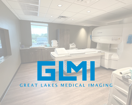
At Great Lakes Medical Imaging (GLMI), we use the latest MRI technology to provide the highest-quality imaging for diagnosing nerve damage and other health conditions. Our advanced machines deliver high-resolution images that help radiologists and doctors accurately identify nerve-related issues.
Experienced Radiologists and Fast Results
Our team of experienced, board-certified radiologists works diligently to interpret your MRI results accurately and efficiently. We understand that timely diagnosis is essential for nerve-related conditions, and we prioritize quick turnaround times to ensure that you get the care you need as soon as possible.
Convenient Locations in Buffalo and Western NY
With multiple locations in Buffalo and Western NY, GLMI offers convenient access to MRI services for residents of the region. We offer flexible scheduling and same-day or next-day MRI appointments for urgent cases.
Take Action Today
If you're experiencing symptoms that may indicate nerve damage, or if your healthcare provider has referred you for an MRI, contact us today to schedule your MRI appointment at Great Lakes Medical Imaging. Our expert team is here to help you get the accurate results and timely treatment you need.
Medical Disclaimer
This article is for general informational purposes only and is not a substitute for professional medical advice. Always consult with a qualified healthcare provider regarding any medical conditions or concerns you may have.
‹ Back